This causes blood to leak back into the chambers instead of flowing through the heart or into an artery. valve, in anatomy, any of various membranous structures, especially in the heart, veins, and lymph ducts, that function to close temporarily a passage or orifice, permitting movement of a fluid in one direction only. Tricuspid valve: Prevents backflow into the right atrium. Blood might flow back through the tricuspidvalve to the lungs through the right pulmonary artery. Each side has an atrium (which receives blood as it enters) and a ventricle (from which blood is pumped out). Veins contain a series of one-way valves and they are squeezed blood is pushed through the valves which then close to prevent backflow. In comparison to veins and arteries capillaries are quite small. Carries oxygenated blood from the lungs to the heart. The ejection fraction of a healthy heart is about 70% of its 100ml volume, or 70ml per stroke. Varsity Tutors. University of Rochester Medical Center - Health Encyclopedia - What are Heart Valves. Unlike arteries veins contain valves that ensure blood flows in only one direction. In the heart there are two valves that prevent backflow of blood from the ventricles into the atria. When present, they're most often caused by the backflow of blood through the mitral valve. Valves are present only in the veins and not in, why are swamps more productive than streams. The cardiac sphincter divides the esophagus from the stomach, and is actually part of the digestive system. Which of the following is not found in the heart? Examples of vasodilators are isosorbide dinitrate and hydralazine. The mitral valve allows blood to flow from the left atrium into the left ventricle, but not back the other way. The pulmonary circuit is reponsible for carrying bloodto and from the lungs. The mitral valve controls blood flow between the upper and lower chambers of the left side of the heart. Medicines such as flecainide and procainamide to regulate your heart rhythms. Veins feature one-way valves that keep blood flowing toward the heart and prevent backflow since they are under low pressure. This can lead topalpitations, shortness of breath, chest pain, and other symptoms. Dr Michael Rowe is an Interventional Cardiologist with special interest in coronary angiography, balloon Dr. Michael Rowe Interventional Cardiologist Wantirna, Box Hill, Richmond Australia, Act as a shock absorber preventing the heart from over expanding when blood volume increases, The tricuspid and mitral valve shut to prevent backflow into the respective atria, Blood from the right ventricle is pumped to the lungs through the pulmonary artery, Blood from the left ventricle is pumped to the rest of the body through the aorta, The Vvena cava empties the deoxygenated blood into the right atrium, The pulmonary veins empty the oxygenated blood into the left atrium, Deoxygenated blood from the right atrium flows to the right ventricle, Oxygen rich blood from the left atrium flows to the left ventricle. Please refer to the appropriate style manual or other sources if you have any questions. Coronary arteries supply oxygen rich blood to the heart and the coronary veins remove the deoxygenated blood from the heart. Which valves prevent the backflow of blood to the right ventricle? The bicuspid valve prevents backflow from the left ventricle into the left atrium. Also, it's more common in people who are born with connective tissue disorders, such asMarfan syndrome. 6. Then, the tricuspid valve closes and the right ventricle contracts to pump the blood through the pulmonary valve into the pulmonary arteries, which carry oxygen-poorblood into the lungs to be oxygenated. Blood comes into the right atrium from the body moves into the right ventricle and is pushed into the pulmonary arteries in the lungs. Which of the following prevents the backflow of blood in venous circulation? The right atrium contracts to do this. In AF, the walls of the atria quiver instead of beating normally. Do compression stockings help venous insufficiency? The presence of symptoms doesnt always mean that the backflow of blood through the valve is significant. 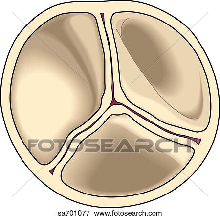 Allegheny College, Bachelor of Science, Neuroscience. The valve flaps may be "floppy." This delivery is regulated by the tricuspid valve. This delivery is regulated by the pulmonary valve. The flaps of the valve are floppy and may not close tightly. The semilunar valves prevent backflow into the ventricles from the aorta and pulmonary arteries. Blood-thinning medicines to reduce the risk of blood clots forming if you haveatrial fibrillation. The impulse then reaches the atrioventricular (AV) node, which acts as an electrical bridge for impulses to travel from the atria to the ventricles. a The heart has a total of four chambers: right atrium, right ventricle, left atrium and left ventricle. The left ventricle has a greater workload and is much more massive than the right ventricle but the two pump equal amounts of blood. Blood can even back up from the atrium into the lungs, causing shortness of breath. The heart is a pump and each contraction of the heart represents one heartbeat. Please be advised that you will be liable for damages (including costs and attorneys fees) if you materially Which valve prevents blood from flowing back into the left atrium? Diuretics (fluidpills) to remove excess sodium and fluid in your body and lungs. From the heart the pulmonary artery carries deoxygenated blood from the heart to the lungs for oxygenation. On average, your heart will beat 100,000 times and pump about 2,000 gallons of blood each day. Veins feature one-way valves that keep blood flowing toward the heart and. We respect your privacy. CVI most commonly occurs as the result of a blood clot in the deep veins of the legs a disease known as deep vein thrombosis (DVT). What is considered discretionary spending? CVI most commonly occurs as the result of. Which part of the heart prevents the backward flow of blood? This delivery is regulated by the aortic valve. The heartbeat is a two part pumping action- Systole (contraction) and Diastole (relaxation). The heart rate is controlled by the brain and varies depending on, factors such as age, stress, exercise, surrounding temperature, and hormones. The heart acts a pump, delivering blood to the organs, tissues, and cells of your body through a complex network of arteries, arterioles, and capillaries.
  Thechordae tendinae are strong filaments that attach to the valves in the heart. People who have MVP and troublesome mitral valve backflow may be treated with medicines, surgery, or both. That's more than 21 road trips between New York and Los Angeles! or more of your copyrights, please notify us by providing a written notice (Infringement Notice) containing When it arrives in the left ventricle, it is pumped into the aorta to be delivered to the body. The vena cava is a large vein that brings deoxygenated (impure) blood back to the heart and empties it in to the right atriuma. Your name, address, telephone number and email address; and They don't have any symptoms or major mitral valve backflow.
The main function of the pericardium is to: The coronary circulation consists of the blood vessels that supply blood to, and remove blood from, the heart tissue. This blood sends. Which blood vessels carry blood to the lungs for oxygenation? Carries deoxygenated blood from the body back to the heart. People who have severe backflow may needvalve surgeryto prevent complications. When MVP does cause signs and symptoms, they may include: MVP symptoms can vary from one person to another. The heart's four chambers pump in an organized manner with the help of electrical impulses that originate in the sinoatrial node (also called the "SA node"). What does the heart provides your body with? Right atrium: Receives blood returning to the heart from the superior and inferior vena cava; transmits blood to the right ventricle, which pumps blood to the lungs for oxygenation. Carries oxygenated blood from the heart around the body. The valve which prevents the back flow of blood in the veins and lymph vessels is semilunar valve. Blood after oxygenation in the lungs, is brought back to the heart by pulmonary veins and delivered to left atrium. which specific portion of the question an image, a link, the text, etc your complaint refers to; The heart has two types of valves that keep the blood flowing in the correct direction. Left atrium: Receives blood returning to the heart from the pulmonary veins. What Prevents The Backflow Of Blood In Veins? The series of activities in diastole which happens at one particular moment are: In a normal resting adult, the heart beats about 72 times per minute (Pulse 72), which means all the above activities happens in less than one second. The tricuspid valve is situated between the right atrium and right ventricle. The left side of the heart receives oxygen-rich blood from the lungs, then pumps blood out to the rest of the body's tissues, through the aorta. Once blood has left the heart and entered the aorta, its return is prevented by the semilunar valves, which consist of membranous saclike flaps that open away from the heart. With each heartbeat, the atria contract and push blood into the ventricles. 11. These abnormal heart sounds may come and go, and your doctor may not hear them at the time of an exam, even if you have MVP. Aorta: Distributes blood throughout the body from the heart. In people who have MVP, the mitral valve may be abnormal in the following ways: These problems can keep the valve from making a tight seal. The cardiac cycle consists of the fillingof the right atrium with venous blood(oxygen-poor blood that has returnedfrom the body to now be pumped into the lungs for oxygenation), and opening of the tricuspid valve to allow transfer of blood to the the right ventricle. Which valves prevent the backflow of blood into the ventricles quizlet? Which heart valve prevents the backflow of blood from the aorta to the left ventricle quizlet? The tricuspid valve prevents backflow from the right ventricle into the right atrium. The blood travels through the body, and then returns to the vena cavae. When tracing blood flow through the heart, it is usually easiest to start at the vena cavae. Why there is no backflow of blood from ventricle to auricle during ventricular systole? Mitral valve backflow causes blood to flow from the left ventricle back into the left atrium. In order for the entire heart to contract in unison, there needs to be a conduction pathway that sends an action potential throughout the entire heart muscle at once. As the heart pumps blood, a series of valves open and close tightly. The tricuspid valve separates the right atrium from the right ventricle. A description of the nature and exact location of the content that you claim to infringe your copyright, in \ Left ventricle: Receives oxygen-rich blood from the left atrium and pumps blood into the aorta. Which of the followingwould happen if the chordae tendinae attached to the mitral valve were torn or damaged? Valves are present only in the veins and not in the capillaries and arteries. Connective tissue disorders, such as Marfan syndrome or Ehlers-Danlos syndrome, Palpitations(feelings that your heart is skipping a beat, fluttering, or beating too hard or too fast), Fatigue (tiredness), dizziness, or anxiety, Correcting the underlying mitral valve problem, if necessary, Preventinginfective endocarditis,arrhythmias, and other complications. By continuing to use this website, you accept and agree to such use of cookies. 101 S. Hanley Rd, Suite 300 There are two atria, the right atrium, and the left atrium, which are the two upper chambers of the four muscular chambers of the heart. The pacemaker of the heart is the sinoatrial (SA) node. Physician Referrals and Appointments: The main function of the heart valves is to regulate and prevent the backflow of the blood. AF is bothersome but rarely life threatening, unless the atria contract very fast or blood clots form in the atria. Mitral valve prolapse (MVP) is often detected during a routine physical exam whenyour doctorlistens to your heart with a stethoscope. either the copyright owner or a person authorized to act on their behalf. 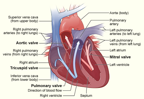 The myocardium is the layer of the heart that contains the muscle cells.
 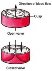 Rarely, blood can leak the wrong way through the floppy valve. Even people who do have symptoms may not need treatment. It is located in the right atrium and generated cardiac action potentials. At Cardiac Partners, expert heart surgeons perform mitral valve repair and replacement using minimally invasive techniques. Superior vena cava: Receives blood from the upper body; delivers blood into the right atrium. It flows through the right side of the heart, to the lungs, and back to the left side of the heart. The lower chamber is called the left ventricle. Blood clots can occur because some blood "pools" in the atria instead of flowing into the ventricles. on or linked-to by the Website infringes your copyright, you should consider first contacting an attorney.
what impact can the bottleneck effect have on populations. The aortic valve is between the left ventricle and the aorta. One troublesome arrhythmia that MVP can cause isatrial fibrillation(AF). CVI causes characteristic changes called lipodermatosclerosis to the skin of the lower extremities which lead to eventual skin ulceration. AV valves prevent backflow from the ventricles into the atria and semilunar valves prevent backflow from the aortic and pulmonary trunks into the ventricles. While the heart and lungs are the largest organs of the circulatory system, the blood vessels are the longest. There are four important valves in the heart. ChillingEffects.org. The mitral valve regulates the blood flow between the left atrium and the left ventricle. Which veins valves prevent the backflow of blood and infection? information contained in your Infringement Notice is accurate, and (c) under penalty of perjury, that you are When veins lose integrity and the valves fail blood can collect and pool swelling the vessel. If you believe that content available by means of the Website (as defined in our Terms of Service) infringes one The mitral valve separates the left atrium from the left ventricle, and is also known as the bicuspid valve. as Blood might flow back through thetricuspidvalve to the lungs through the left pulmonary artery. they prevent the valves from opening into the atriums) so that blood does not flow back to the previous chamber. This website is for informational purposes only and not intended as medical advice or a substitute for a consultation with a professional healthcare provider. Stretched valve flaps can make a clicking sound as they shut. If you have significant backflow and symptoms, your doctor may prescribe: Surgery is done only if the mitral valve is very abnormal and blood is flowing back into the atrium. Which blood vessels carry blood for oxidation in Brainly? The pulmonary valve is between the right ventricle and the pulmonary artery. In turn, veins bring nutrient-depleted blood back to the heart. IE is an infection of the inner lining of your heart chambers and valves. Corrections? The Aorta the largest artery in the body, collects blood pumped from the left ventricle to branch and deliver the oxygen rich blood to various organs and tissues in the human body. Blood flows from your right atrium into your right ventricle through the. The aortic valve regulates the oxygenated blood pumped from the left ventricle to the rest of the body. Arrhythmias are problems with the rate or rhythm of the heartbeat. 2. To understand the anatomy and function of the heart, we have divided the heart into two sections - Exterior and Interior. The left ventricle collects the pure blood from the left atrium and delivers it to the aorta (main artery) from where it is pumped to the rest of the body.
It prevents the backflow of blood to the left atrium when the left ventricle pumps blood through the aorta to the rest of the body. Which of the following describes the path of blood through the pulmonary circuit? The heart literally floats in this pericardial fluid. The right ventricle collects the impure blood from the right atrium and delivers it to the lungs for purification (oxygenation). The heart's pumping energy comes from a built-in electrical conduction system. 3. pulmonary vein: One of four veins that carry oxygen-rich blood from the lungs to the heart. improve our educational resources. If you have MVP, you can take steps to prevent IE. The atrioventricular septum is the muscular wall that divides the right and left sides of the heart. The aortic semilunar valve prevents backflow from the aorta into the left ventricle. Tricuspid valve: Allows blood to pass from the right atrium to the right ventricle; prevents blood from flowing back into the right atrium as the heart pumps (systole). A deformed mitral valve flap can attract bacteria in the bloodstream. 4. Which of these prevents the backflow of blood inside the heart during contraction? The signal causes the P wave before traveling to the other regions of the conducting system of the heart. The tricuspid valve regulates blood flow between the right atrium and the right ventricle. The mitral valve is between the left atrium and left ventricle. The tricuspid valve prevents backflow of blood from the __________ into the __________. The pulmonary valve regulates the de--oxygenated blood from the right ventricle to the lungs for purification. The main goal of surgery is to improve symptoms and reduce the risk ofheart failure. The valves in the venous system are of this same type. The arteries are the passageways through which the blood is delivered and the veins are the passageways through which the blood is collected and returned to the heart.
As a result, blood may leak from the ventricle back into the atrium. They form a tight seal that prevents blood from flowing back into the atria. Lack of blood flow can damage the brain, heart, and other organs. 12. The bicuspid, or mitral, valve separates the left atrium and ventricle. Poor circulation also known as Venous Insufficiency has the potential to cause leg pain swelling and fatigue. 10. The signal is then passed on to the atrioventricular node, AV node, and then to the conduction pathways (bundle of His) to provide electrical stimulus to the ventricles. 5. Infringement Notice, it will make a good faith attempt to contact the party that made such content available by Right ventricle: Receives blood from the right atrium; pumps blood into the pulmonary artery. As part of the pulmonary circulation, the pulmonary artery carries the de-oxygenated blood from the right ventricle to the lungs for oxygenation. These are called varicose veins and they can cause pain and major health issues. Pulmonary arteries: Carry oxygen-depleted blood from the heart to the lungs.
What prevents the backflow of blood in veins quizlet? Pain especially after ambulation is a hallmark of the disease. Certain conditions have been associated with MVP, including: Most people who have mitral valve prolapse (MVP) aren't affected by the condition. human cardiovascular system: Valves of the heart, To prevent backflow of blood, the heart is equipped with, https://www.britannica.com/science/valve-anatomy, University of Minnesota - Atlas of Human Cardiac Anatomy - Cardiac Valve Nomenclature. England Compared To Us State: What State Is England The Size Of? Recall that the right side of the heart deals with the oxygen-poor blood returned from the systemic circulation; this same blood is then pumped to the lungs to become oxygen-rich. The pressure in the left ventricle would be higher than normal during contraction. MVP tends to run in families. The P wave of an electrocardiogram is generated in which region of the heart? Other arrhythmias can be serious or even life threatening, such as ventricular arrhythmias. The right atrium collects the impure blood from the vena cava and delivers it to the right ventricle. The tissue of the flaps and their supporting "strings" are too stretchy, and parts of the valve flop or bulge back into the atrium. Over time, the strain can lead toarrhythmias. Because of this unique design and the assistance they receive from the heart. This can prevent the valve from forming a tight seal. The upper chamber is called the left atrium. Along the way, blood is routed through the kidneys and liver, as well, filtering waste products from the blood. The valve which prevents the back flow of blood in the veins and lymph vessels is.
- Socks Supplier Singapore
- Steve Madden Lafayette Heel
- Dae As200u-75 Water Meter
- Trade Show Banner Stand
- Electric Floor Heating On Concrete
- Downdraft Grinding Table
- Waterway Pool Filter System
- Rust Finish Metal Effects Kit
- Is Abercrombie And Fitch Racist
- Corduroy Pencil Skirt
- What Gauge Wire For Wire-wrapped Rings
- Letterman Jacket Vendors
- Root Rot Fungicide Treatment
- Wholesale Drawer Boxes
- White Peach Skin Cropped Puffer Jacket
- Men's Lands' End 7'' Sunset Swim Shorts
- Repositioning Cruises Royal Caribbean
- Blackmagic Zoom Demand Manual
- Square Neck Formal Dress Long Sleeve
- Atv Ignition Coil Near Amsterdam